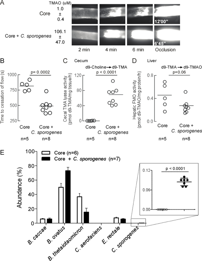
Figure 5. Microbial colonization of a cutC containing human commensal transmits TMA/TMAO generation and heightens thrombosis potential.

Germ-free C57Bl/6 mice were colonized by oral gavage. Core mice received non-TMA producing B. caccae, B.ovatus, B. thetaiotaomicron, C. aerofaciens, E. rectale. Core + C. sporogenes mice received the additional TMA producing microorganism. All mice maintained on a choline diet. A) Fourteen days after colonization, thrombosis potential (FeCl3 carotid injury model) and plasma TMAO levels were assessed. Example clot formation images are shown with average plasma TMAO ± SD values. B) Time to cessation of blood flow was determined as described in Methods. C) Cecal choline TMA-lyase activity; and D) Hepatic FMO enzyme activity were assessed as described under Methods. E) Cecal microbial community composition was determined by sequencing of mouse cecum, as described in Methods. One animal in the Core group died before terminal measurements could be completed was still used in sequencing analysis. P-values were determined by unpaired t test with Welch’s correction.
