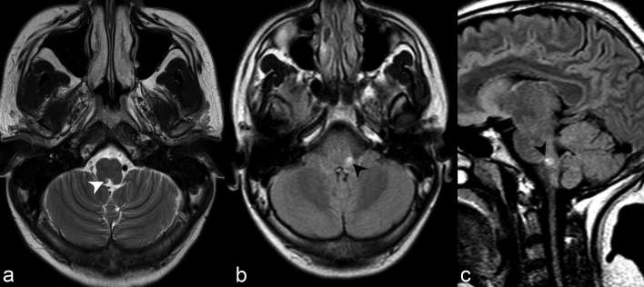
Figure 3.
(a) Axial T2 weighted imaging at the level of the medulla demonstrates abnormally increased signal in the right dorsomedial medulla adjacent to the fourth ventricle in the region of the area postrema (white arrow head). The lesion was poorly visualized on FLAIR, partly secondary to the decreased sensitivity of posterior fossa lesions on such sequence. Axial (b) and sagittal (c) FLAIR of a different patient with hiccups related to area postrema syndrome demonstrate abnormally increased signal in the left dorsal medulla (black arrow heads). These lesions are often most specific for NMOSD and lesions correspond to areas of high aquaporin-4 (AQP4) expression. AQP4, high aquaporin-4; FLAIR, fluid attenuation inversion recovery; NMOSD, neuromyelitis opticaspectrum disorder.

