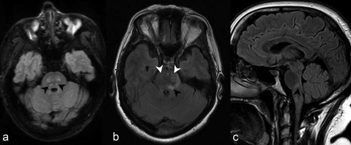
Figure 4.
Axial 3D and 2D FLAIR at the level of the superior cerebellar peduncles (a, b) and sagittal FLAIR (c) of three different patients demonstrate foci of increased signal in the dorsal pons regional to the fourth ventricle (black arrow heads). Note the additional involvement of the CST more anteriorly with the dispersed, linear pyramidal tract bundles discretely outlined by abnormally increased signal (white arrow heads) (discussed later). 2D, two-dimensional; 3D, three-dimensional; CST, corticospinal tract; FLAIR, fluid attenuation inversion recovery.

