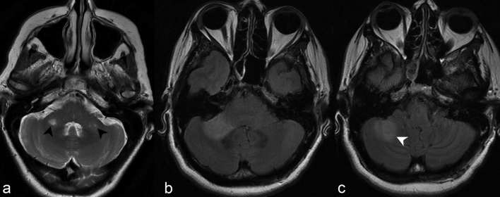
Figure 5.
(a) Axial T2 weighted imaging at the level of the cerebellopontine angle demonstrates discrete foci of abnormally increased signal in both middle cerebellar peduncles (black arrow heads). (b) Axial FLAIR at the same level in another patient demonstrates a confluent focus of abnormally increased signal in the right middle cerebellar peduncle. (c) At a slightly more caudal level, the abnormal signal extends posterolaterally to involve the right cerebellar hemisphere (white arrow head). FLAIR, fluid attenuation inversion recovery.

