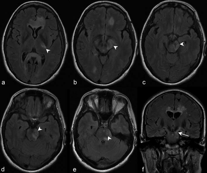
Figure 6.
Axial FLAIR images demonstrate a contiguous region of abnormally increased signal affecting the left CST (white arrowheads), from the posterior limb of the left internal capsule (a), to the cerebral peduncle (b–d) and pontine pyramidal tract bundles (e). Note the additional full-thickness involvement of the splenium of the corpus callosum, which is often termed “arch bridge pattern” (a, black arrows) (discussed later), and confluent involvement of the genu beginning to extend into the cerebral white matter (a, black arrow head). Coronal FLAIR imaging of the same patient at the level of the third ventricle (f) depicts the contiguous involvement of the left CST extending from the posterior limb of the internal capsule caudally to the cerebral peduncle (white arrow). Note again the extension of the signal abnormality from the corpus callosum into the left cerebral white matter (f, black arrow head). CST, corticospinal tract; FLAIR, fluid attenuation inversion recovery.

