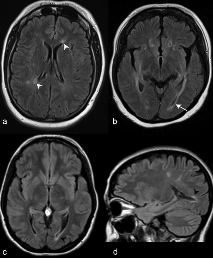
Figure 8.
(a) Axial FLAIR imaging at the level of the corona radiata demonstrates increased periependymal signal abnormalities adjacent to the frontal horn and body of the left and right lateral ventricles, respectively (white arrow heads). (b) Axial FLAIR imaging at the level of the hypothalamus in a different patient demonstrates periependymal signal abnormality adjacent to the atrium of both lateral ventricles. Note the posterior extension of the left periependymal lesion to involve the periependymal white matter adjacent to the left occipital horn (white arrow). Axial (c) and sagittal (d) FLAIR of a third patient similarly demonstrate a periependymal lesion adjacent to the left occipital horn of the lateral ventricle (black arrow heads). FLAIR, fluid attenuation inversion recovery.

