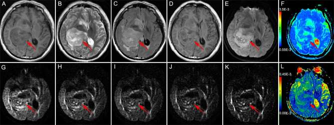
Figure 2.
Brain MRI of a 30-year-old male with Grade II astrocytoma in the right dorsal thalamus The solid part of the tumor (the red arrow head) is hypointense on T1WI (a) and shows mild hyperintensity on T2WI (b) and T2FLAIR (c); the solid part of the tumor shows mild enhancement on enhanced T1WI (d). The DWI (b = 1000 s mm–2) shows the solid part of the tumor as hyperintense (e); ADC value was 0.926 × 10⁻³ mm2 s–1 (f); the DWI [b = 1800 s mm–2 (g), b = 2500 s mm–2 (h), b = 3000 s mm–2 (i), b = 3500 s mm–2 (j), b = 4000 s mm–2 (k)] showed the solid part of the tumor as hypointense; the UHBV-ADC value was 0.0899 × 10⁻³ mm2 s–1 (l). ADC, apparent diffusion coefficient; T1WI, T1 weighted image, T2WI, T2 weighted image; T2 FLAIR, T2 fluid-attenuated inversion recovery image; UHBV-ADC, ultra-high-b-value-ADC.

