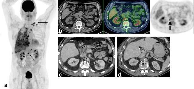
Figure 1.
A 70 year-old male with epithelioid malignant pleural mesothelioma. (a) Maximum intensity projection FDG-PET image shows extensive right pleural malignancy and metastatic adenopathy in the mediastinum and left supraclavicular fossa (arrow). Further metabolically active nodes are seen below the diaphragm (dotted arrow). (b) PET/CT image (CT, left; fused PET/CT, middle; PET, right) shows two foci of abnormal FDG uptake in tiny retrocaval lymph nodes. (c) Concurrent contrast-enhanced CT shows normal sized retroperitoneal nodes, with no size significant lymphadenopathy. (d) Follow-up contrast–enhanced CT performed 7 weeks later shows interval development of enlarged lymph nodes in same location, indicating progressive metastatic disease and confirming baseline PET/CT findings. FDG, fludeoxyglucose.

