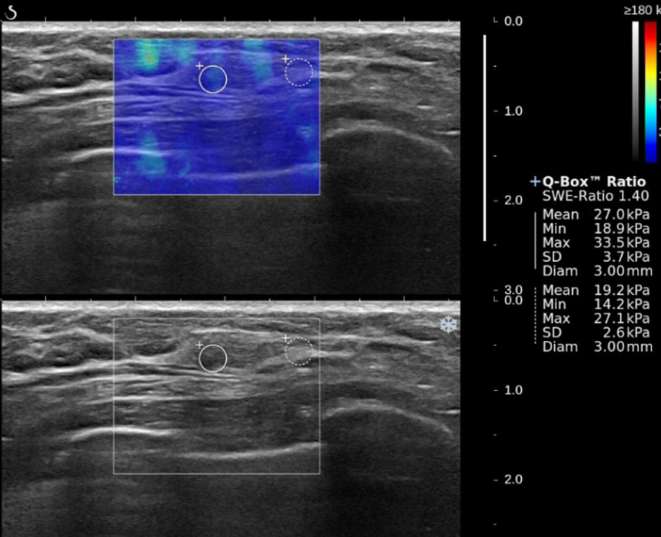
Figure 2.
A 51-year-old female with a 4 mm, Grade 1, ductal carcinoma in situ in the left breast. B-mode ultrasound (bottom) shows that mass has irregular shape and non-circumscribed margin. SWE color map (top) shows that blue color around the lesion continues vertically on the cutaneous side (Pattern 2) and low SWE values (Emax, 33.5 kPa; Emean, 27.0 kPa; Eratio, 1.4; SD, 3.7 kPa). On pathology, the cancer showed no lymphovascular invasion, negative ER, negative PR, negative HER2, and negative Ki-67.

