
Figure 3.
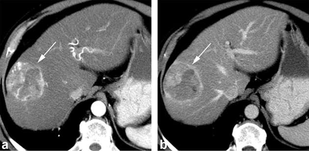
Axial contrast-enhanced CT obtained during the arterial (a) and portal venous (b) phase shows a 5.6 cm hepatocellular carcinoma diagnosed at liver resection with 50% FAI score. The lesion shows arterial phase hyperenhancement (arrow, a), washout and capsule appearance on the portal venous phase image (arrow, b) and mosaic architecture. The lesion was categorized as LR-5. FAI, fractional allelic imbalance.