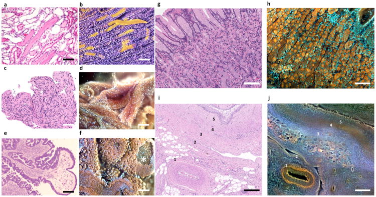
Figure 6. Additional shape and colour information available in MUSE vs. conventional H&E images.
a–f, Enhanced surface shape information. a, Standard FFPE-H&E histology of human kidney with proteinaceous casts filling some tubules. b, Same kidney imaged with MUSE, and pseudocoloured for clarity. Casts can be seen as cylindrical structures, including one that was dislodged from its original site. c, H&E Schwannoma. Swirling nature of this tumor can be appreciated. d, MUSE of same specimen, in which the 3D organization of the tumor can be appreciated. e, Seromucinous ovarian carcinoma, H&E. Carcinoma-lined tufts surround fibrovascular cores are visible. f, Same tumor imaged with MUSE, revealing cauliflower floret-like clumps of malignant epithelial cells outside fibrovascular cores. A window into a core is visible, top right.
g–j, Broader colour gamut reveals novel tissue contrast. g,h, Corresponding regions of normal human stomach fundus and i,j, porcine renal pelvis are shown, with MUSE specimens stained using rhodamine and Hoechst. g, Conventional FFPE H&E; h, MUSE, fluorescence image from a similar region. The chief (brown) and parietal cells (orange) are much easier to distinguish in the fluorescence MUSE image. i, Fixed porcine renal tissue, H&E; j, corresponding region, MUSE fluorescence mode. Stromal features, some identified by number, are easier to distinguish in the MUSE image vs. H&E. Scale bar = 100 μm.

