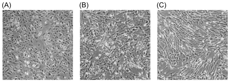
Figure 1. PEL cells induce a myofibroblastic morphology in mesothelial cells.
Phase-contrast images of a primary culture of normal human mesothelial cells (A) showing a cobblestone-like morphology. Transition to a myofibroblastic morphology, characterized by elongated, spindle shaped cells, and a crisscross pattern of growth, is induced after co-culture with PEL-derived cell lines. Mesothelial cells are shown after 4 (B) and 8 (C) days of co-culture with CRO-AP/2 cells. This morphological change is consistent with the induction of epithelial-mesenchymal transition by the TGF-beta released by PEL cells.50

