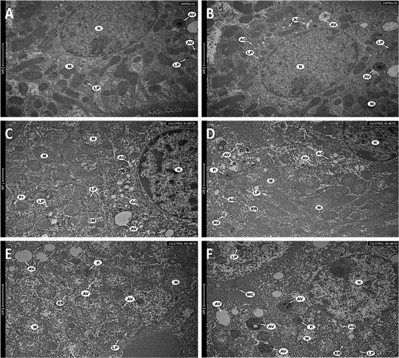
Figure 3. Fragments of the kidney cells (A, B) of an untreated animal, (C, D) of a hamster treated with 50 nM, (E, F) of a hamster treated with 200 nM.
Nucleus (N), mitochondria (M), single autophagic vacuoles (AV), primary lysosomes (LP), Golgi apparatus (AG), polyribosomes (Pr), cell membrane (CM), endoplasmic reticulum (ER), peroxisomes (P.) and microvilli (MC). Magnification 8 200× (A, B, D–F), 9 900× (C).

