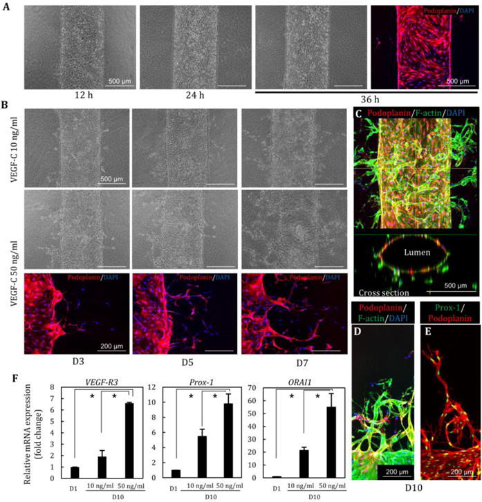
Fig. 2. Lymphatic vessel structures and lymphangiogenesis induced by VEGF-C.

(A) Lymphatic endothelial cells (LECs) were adhered to microchannels in 12 h, resulting in the formation of the lymphatic vessel in 24–36h. (B) Lymphatic morphogenesis was induced in a dose-dependent manner of VEGF-C (10, 50 ng/ml) at D3, D5, and D7. Podoplanin was expressed in lymphatic angiogenic sprouting. (C) Lumen structure of lymphatic vessel after 10 days of perfusion culture. (D, E) Lymphatic sprouting and single cell migration stained by podoplanin and Prox-1. (F) Relative gene expression changes at D1 and D10 (VEGF-C 10 and 50 ng/ml). High concentration of VEGF-C induces increasing the expression of VEGF-R3, Prox1, and ORAI1. *, p<0.01, error bar=±SD.
