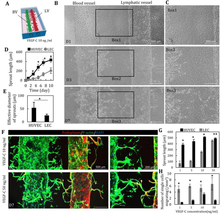
Fig. 3. Lymphatic and blood vessel structures in a collagen matrix.

(A, B) LA and VA were induced by the addition of VEGF-C (10 ng/ml). HUVEC started to migrate and sprout into collagen gel at D3, prior to LEC sprouting. (C) Quantitative comparison of sprouting length (D) and effective diameter (E) of HUVEC and LEC. n=12. LA lagged behind VA. (F) Immunostaining of podoplanin (red) and F-actin (green) and nuclear (blue) of LA and VA at D7 at 10 and 50 ng/ml of VEGF-C. (G) Enhancement of LA and VA at D7 in a dose-dependent manner of VEGF-C (1, 5, 10 and 50 ng/ml). n=5. (H) Single cell migration of HUVEC and LEC at D7. n=5. *, p<0.01; **, p<0.05, error bar=±SD.
