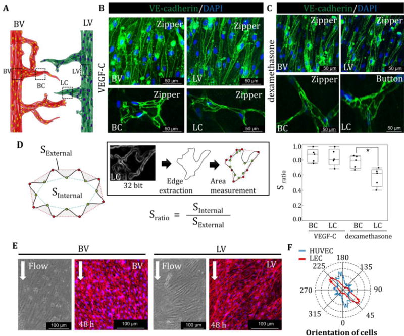
Fig. 4. Cell-cell junctions in blood and lymphatic vessels and orientation of cells.

(A) VE-cadherin staining in lymphatic vessel (LV), blood vessel (BV), blood capillaries (BC), and lymphatic capillaries (LC). (B) VE-cadherin was highly expressed at the boundary of cells in the presence of VEGF-C in BV, LV, BC, and LC, forming “zipper-like junctions”. (C) VE-cadherin expression was localized at the cell-cell boundary with dexamethasone only with the addition of VEGF-C at BV and LV. Zipper-like structures formed at BC. In contrast, LC start to take on an “oak-leaf” appearance, resulting in the formation of button-like structures in LC. (D) Sratio is defined by the internal area of cells divided by the external area of cells. LEC in LC formed serrated structures compared to VEC in BC in the presence of dexamethasone. (E) Representative images of cell orientation in the BV and LV and quantification of directionality by FFT (F). *, p<0.01, error bar=±SD.
