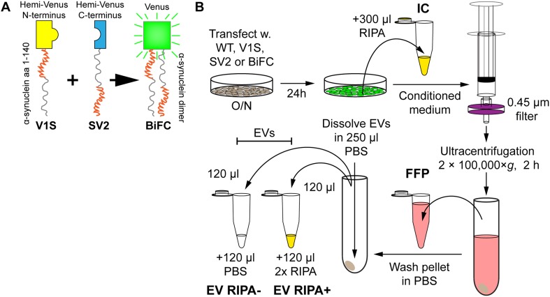
Fig. 1.
Preparation of cell-derived samples for the study of α-syn secretion. a In addition to human WT α-syn, the V1S (yellow) and SV2 (blue) constructs (α-syn fused with either half of Venus) were used. Also, V1S and SV2 were co-transfected in a BiFC. The N-terminal region of the α-syn portion is shown in red, whereas the C-terminal region is shown in gray. Upon α-syn dimerization of V1S and SV2, the protein aggregate fluoresces (green). b SH-SY5Y cells were transfected overnight. The cells were washed once in medium, followed by incubation for 24 h. The ensuing medium was collected, filtered to remove dead cells and debris, ultracentrifuged, upon which the transfected cells were washed in PBS and lysed in RIPA for the IC fraction. The medium supernatant, from the ultracentrifugation (FFP) was saved, after which the pellet was washed once followed by an exchange of tubes before the second ultracentrifugation. The ensuing pellet was reconstituted in PBS, split in two before adding either 2 × RIPA at a 1:1 ratio (EV RIPA+) or additional PBS at a 1:1 ratio (EV RIPA−), to get the two EV fractions

