
Fig. 5.
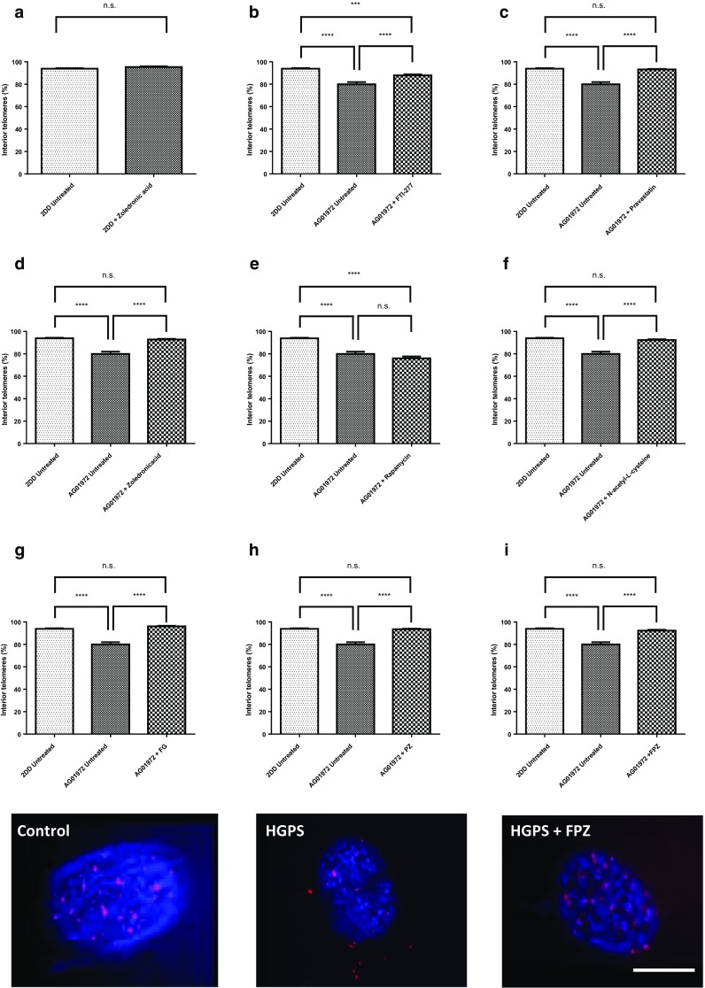
Comparisons of the fraction of telomeres found in the residual nuclei of DNA Halo preparations of HGPS before and after drug treatments. Images of fixed control and HGPS DNA halos stained with both DAPI (blue) and Cy3 (red) telomeric PNA probes were captured at ×100 magnification. Residual nuclei were delineated and the fraction of telomeres determined inside and outside of the residual nuclei. Results for the treatments are shown as a 2DD with zoledronic acid, b FTI-277, c pravastatin, d zoledronic acid, e rapamycin, f N-acetyl-l-cysteine, g FTI-277 and GGTI-2133, h pravastatin and zoledronic acid and i FTI-277, pravastatin and zoledronic acid. The proportion of interior telomeres is expressed as a percentage of the total number of telomeres present. Error bars represent ± SEM. Scale bar = 10 µM