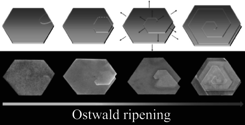
Fig. 6.

Schematic illustration of growth of the multi-layered spiral nanoplate (the upper row) and the corresponding SEM images (the lower row). From left to right are morphologies of different growth stages and gradually matured. Red arrows stand for the growth direction
