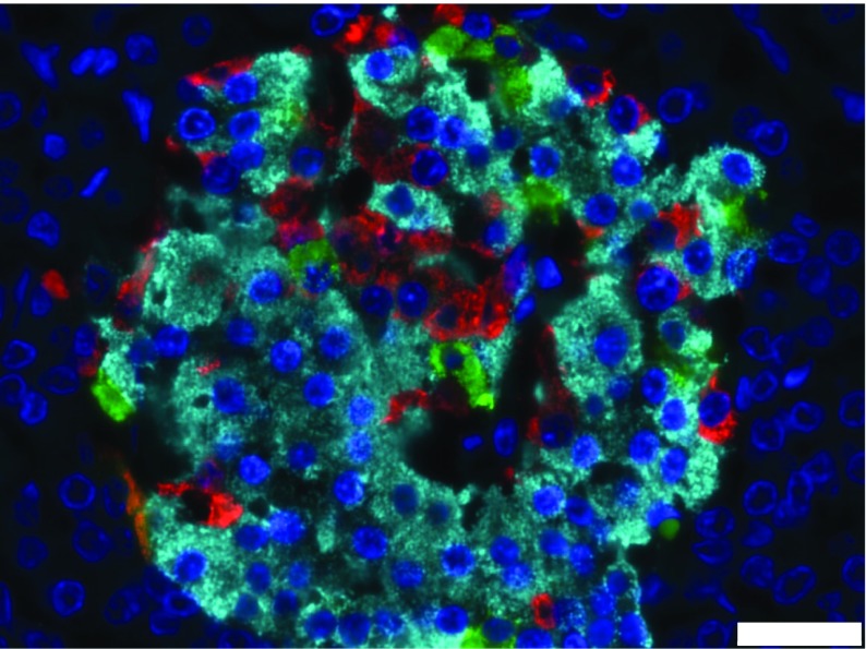
Fig. 3.
Cellular composition of a human islet of Langerhans from a healthy individual. Individual endocrine cell subtypes were identified by immunofluorescent analysis after staining with antisera directed against insulin (light blue), glucagon (red) and somatostatin (green). Cell nuclei were stained with DAPI (dark blue). Scale bar, 25 μm. The image was captured in our laboratory in Exeter and is from a case held in the EADB collection. This figure is available as part of a downloadable slideset

