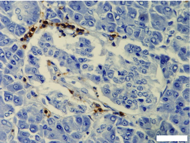
Fig. 4.
Example of an inflamed islet from a child newly diagnosed with type 1 diabetes. Lymphocytes were immunostained in brown with an antibody directed against CD45. Small numbers of lymphocytes are found within the core of the islet but most are located peripherally, with the majority focused at one pole of the islet. Scale bar, 20 μm. The image was captured in our laboratory in Exeter and is from a case held in the EADB collection. This figure is available as part of a downloadable slideset

