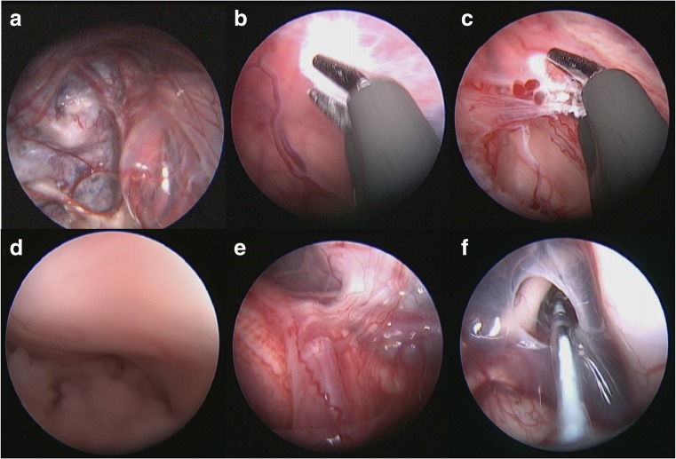
Fig. 3.
Intraoperative imaging demonstrating the inferior aspect of the cyst bulging up into the cavity a; cyst wall being coagulated b; choroid plexus lining cyst wall c; right foramen of Lushka d; lateral aspect of brainstem with the right lower cranial nerves and vertebral artery/PICA seen e; fenestration into pre-pontine cistern f

