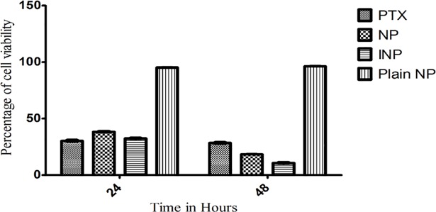
Fig 7. Cell viability studies of NP and INP, PTX and Plain NP, incubated with MDA-MB-468 breast cancer cells for 24 and 48 hours.
“The cell viability was studied by MTT assay using 96 well plates and values were compared with blank nanoparticles and control (PTX solution Analysis of variance (ANOVA) and linear regression analysis was performed on the tumor volume. Means (n = 3) values were compared with two way ANOVA test. Means (n = 3) values were compared with two tailed student t test. NPs and INP showed statistical differences of ***P<0.001 as considered more significant than PTX”.

