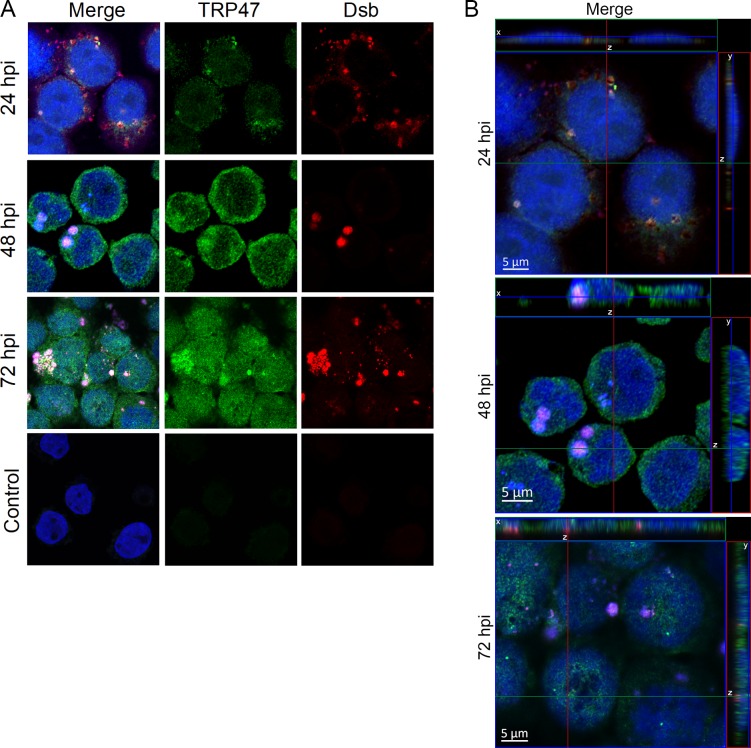
Fig 1. TRP47 localizes to the nucleus of E. chaffeensis-infected cells.
(A) E. chaffeensis-infected and uninfected control THP-1 cells were fixed and probed with mouse anti-TRP47 (green), rabbit anti-Dsb (red), and DAPI (blue), and visualized by scanning confocal laser microscopy. For each timepoint post-infection, one representative optical slice from the z stack is shown. TRP47 was detected in all three timepoints in the infected cells. TRP47 was mainly found in cytoplasm at 24 hpi (in and around the morulae), whereas increased TRP47 was detected in the nucleus at 48 and 72 hpi. Ehrlichial Dsb (red) in micrographs confirmed presence of E. chaffeensis in infected cells at 24, 48 and 72 hpi. We did not observe staining for TRP47 or Dsb in uninfected control. (B) Orthogonal projections of optical slices from a z-stack confirmed the presence of TRP47 in the nucleus especially at 48 and 72 hpi. Top panels show an x-z projection and right panels show a y-z projection. The positions of the x and y axes within the projections denote the z depth of the slice shown in the center.

