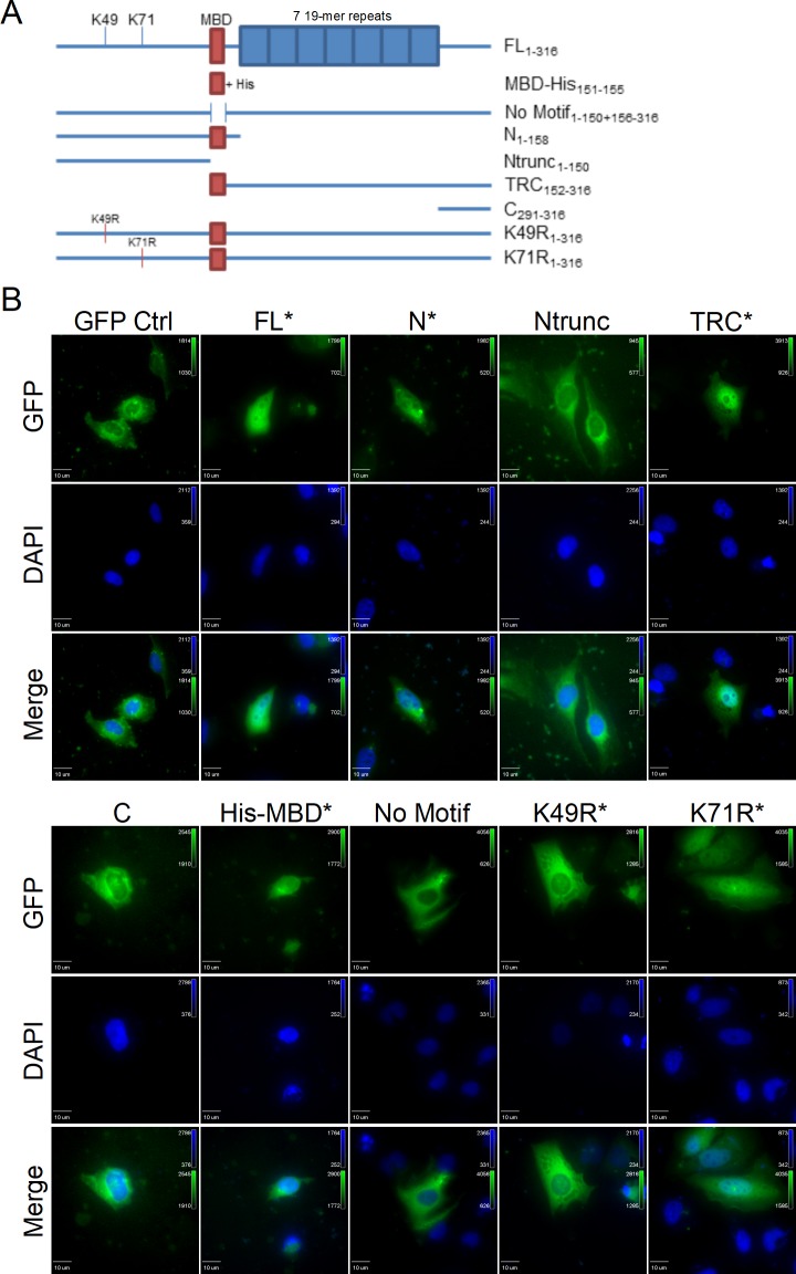
Fig 2. A MYND-binding domain is responsible for TRP47 nuclear translocation.
(A) Schematic showing GFP-tagged TRP47 expression constructs. Red boxes indicate presence of the MYND-binding domain (MBD). Blue boxes represent the seven 19-mer repeats of the TRP47 tandem repeat region. Open space between two lines indicates a deletion mutation. Subscripts on construct labels indicate which amino acid residues are included. Constructs shown are full-length (FL), His-tagged MBD (MBD-His), full-length TRP47 without the MBD (No Motif), N-terminal (N), truncated N-terminal without the MBD (Ntrunc), tandem repeat-C-terminal overlapping the MBD (TRC), C-terminal (C), K49R mutant (K49R), and K71R mutant (K71R). (B) Ectopically expressed GFP-tagged constructs fluoresce green and DAPI-stained nuclei fluoresce blue. The FL, N, TRC, His-MBD, K49R, and K71R constructs contained the MBD (marked by asterisks), while the Ntrunc, C, and No Motif constructs did not. The FL and His-MBD TRP47 constructs exhibited strong nuclear localization and the N, TRC, and K71R TRP47 constructs exhibited nuclear and diffuse cytoplasmic localization. Exclusively cytoplasmic localization was observed with the Ntrunc, C, No Motif, and K49R mutant TRP47 constructs. The GFP negative control construct was also observed only in the cytoplasm. Due to differences in expression levels of the constructs, different exposure times were used to image each construct.

