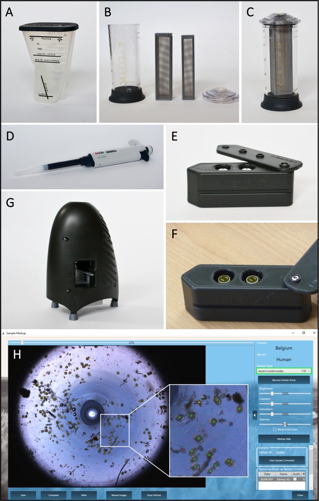
Fig 1. Materials needed to perform the FECPAKG2 method.
(A) The FECPAKG2 sedimenter with indications of the water line and the saline lines used to analyse faeces of cattle or sheep and horses. For analysis of human samples, the saline line for sheep and horses is applied. (B) The filtration unit with cylinder base, cylinder top and two filter frames. (C) The unit fits together and can be disassembled for cleaning. The two filter frames have different mesh sizes (425μm and 250μm) and are designed to be used together. Samples are collected from the filtration unit using a pipette with special extension tip designed to fit into the filtration unit and the top of the cassette (D). The FECPAKG2 cassette with its two counting wells with central light rods is shown in side view (E) and from the top containing a sample (F). A filled cassette is fed into a portal of the MICRO-I device (G) and then pulled through automatically for imaging of each well. (H) A standard image that is produced by the FECPAKG2 and presented to the user. Some debris and numerous STH eggs are visible. Visible Ascaris lumbricoides eggs (n = 118) are marked with green dots, the Trichuris trichiura eggs are marked with yellow dots (n = 29) and the hookworm eggs are marked with a red dot (n = 5). After mark-up, this information is stored and remains available for review by authorized users.

