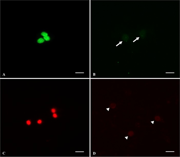
Fig 8. Representative dead cells and fragmented DNA on testicular tissue from prepubertal cats after warming, 24 h, and 5 days of in vitro culture.
(A) live cells with fragmented DNA showing bright green nucleus, (B) live cells with intact membrane (white arrows), (C) dead cells showing bright red nucleus, (D) live cells (white arrow heads). Bar = 5 μm.

