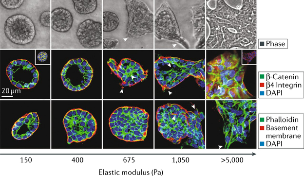
Fig. 2 |. Matrix stiffness regulates the epithelial-to-mesenchymal transition.
Phase contrast and fluorescent images of mammary epithelial ceLL colonies on polyacrylamide hydrogels of indicated stiffness (150–5,000 Pa) with Matrigel overlay are shown. Microscopy images show colony morphology after 20 days. The fluorescent images show β-catenin (green) before and after (inset) triton extraction, β4 integrin (red), epithelial cadherin (E-cadherin) (red; inset) and nuclei (blue). In the bottom images, actin (green), laminin 5 (basement membrane; red) and nuclei (blue) are shown. DAPI, 4’,6-diamidi- no-2-phenylindole. Figure is reproduced with permission from REF.3, Elsevier.

