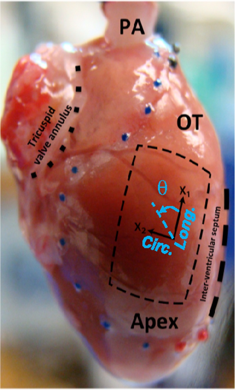
FIG. 2.

Isolated rat heart and right ventricular free wall (RVFW), denoted by a square slab (crossed lines), and the coordinate basis {x1,x2} used for histological measurements. The directions x1 and x2 approximately represent the longitudinal (apex-to-outflow tract) and circumferential directions, respectively. Note that the transmural direction is perpendicular to the x1-x2 plane. The angle θ (positive anticlockwise) denotes the orientation of myo- and collagen fibers (denoted by θm and θc, respectively) in the x1-x2 plane. Reproduced with modification with permission from Valdez-Jasso et al., J. Physiol. 590, 4571-4584 (2012). Copyright 2012 John Wiley and Sons.
