Figure 3.

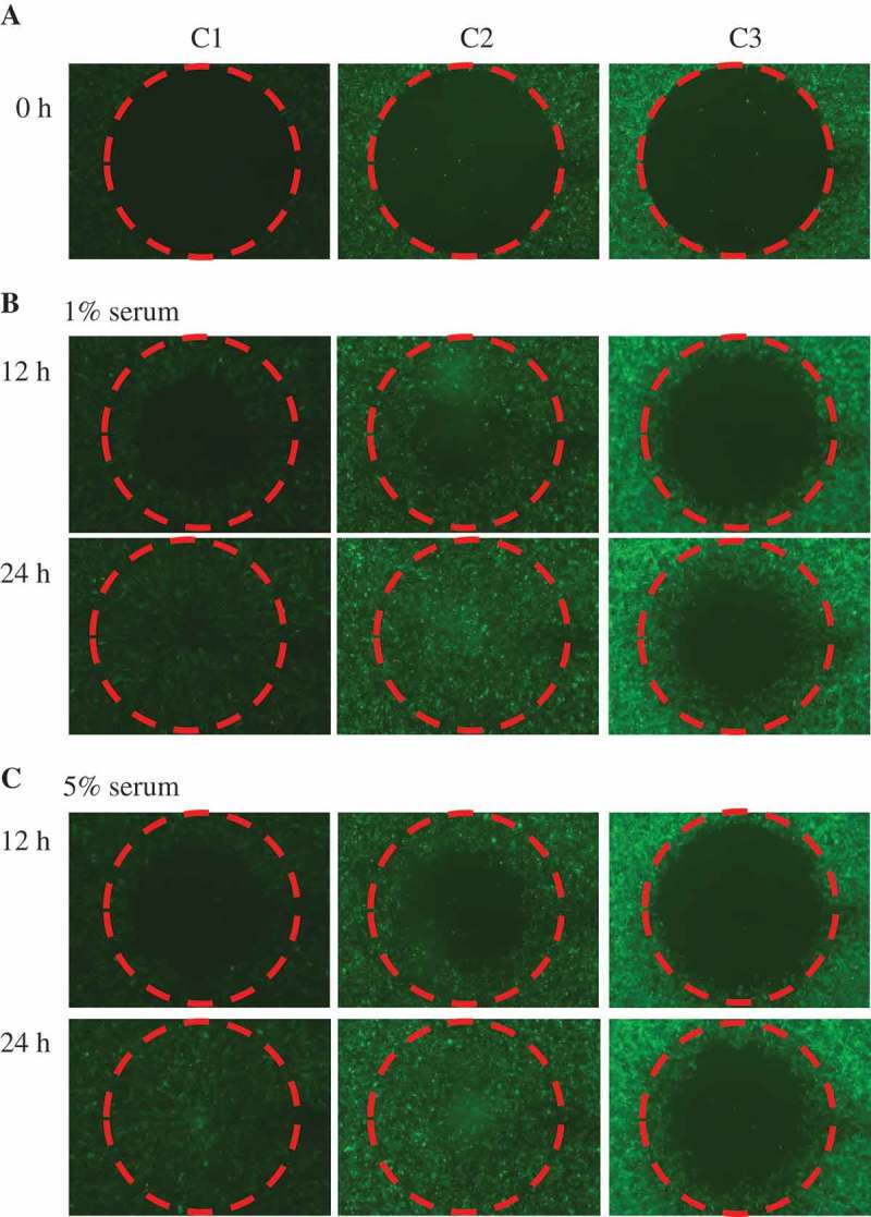
MCF10A.pCDH co-cultured with MCF10A.NeuT cells show enhanced migration ability compared to MCF10A.NeuT cells.
(a) Cells were seeded and grew into 100% confluence. The stoppers were removed at time zero (0). (b) C2 cells migrate similarly to C1 cells but faster than C3 cells 12 hrs and 24 hrs after in the presence of 1% serum. (c) Cells were cultured in medium supplemented with 5% serum.
