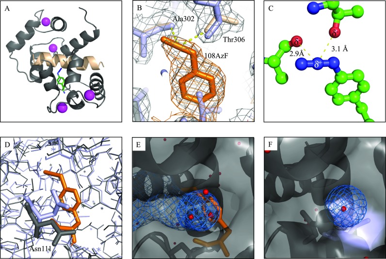
FIG. 4.
Crystal structure of the CaM108AzF+P2 complex. (a) Overall structure in cartoon representation. CaM is colored grey with 108AzF in green/blue. The peptide is colored in wheat and Ca2+ ion in magenta. (b) Polder OMIT map for 108AzF contoured at 3σ illustrating clear electron density for the incorporated amino acid in orange with an additional 2Fo-Fc map shown in grey and the wild-type structure (pdb:1cdm) superimposed in light blue. (c) Details and distances of the weak dipole-dipole interactions between the azido group of 108AzF and carbonyl groups of Ala302 and Thr306 of the binding peptide. (d) Superposition of the CaM108AzF+P2 complex (grey with AzF in orange) with pdb:1cdm (light blue) illustrating the different orientation of Asn111. (e) Representation of the CaM108AzF+P2 complex illustrating the solvent accessibility of 108AzF (orange) due to the orientation change of Asn111. Water molecules are shown in red, and the solvent accessible cavity is shown as blue mesh. (f) Representation of pdb:1cdm at Val108 (light blue) reveals the lack of the solvent accessible cavity in the wild-type complex.

