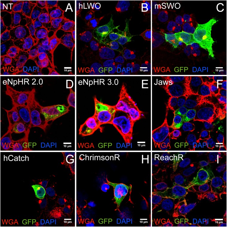
FIGURE 1.
Optogenes and their trafficking profile in transfected HEK cells. HEK293 cells were labeled with membrane marker WGA-Rhodamine and anti-GFP antibodies. Representative confocal images from non-transfected control (NT) (A), vertebrate opsins human long wavelength opsin (hLWO) (B) and mouse short wavelength opsin (mSWO) (C) and microbial opsins eNpHR 2.0 (D) and eNpHR 3.0 (E), Jaws (F), hCatCh (G), ChrimsonR (H), and ReaChR (I). Scale bar: 10 μm.

