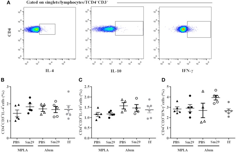
Figure 2.
Profile of TCD4 subsets in animals immunized with Sm29 Alum and Sm29 MPL immunization. The percentage of CD4+IL-4+, CD4+IFN-γ+ and CD4+IL-10+ cytokines was evaluated by TCD4 intracellular staining in spleen cells from 5 to 6 animals of each group. Intracellular cytokine staining was performed in the absence of polyclonal or antigen specific stimulated to assesses differentiation induced by the vaccine formulation in vivo. Data analysis was carried out as demonstrated in (A) within singlet cells/lymphocyte region, cells expressing CD4 and CD3 molecules were selected and the percentage of CD4+ cells producing IL-4 (B), IL-10 (C) and IFN-γ (D) was evaluated in MPLA (closed triangles), Sm29 MPLA (closed circles), Alum (open triangles), Sm29 Alum (open circles) and infected/treated (gray circles) groups. Results are representative of one from two independent experiments. No significant differences between groups were observed.

