Fig. 3.
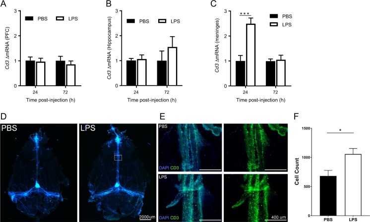
T lymphocytes transiently accumulate in the meninges prior resolution of depression-like behavior. a Cd3 mRNA level in the prefrontal cortex (PFC) 24 h and 72 h after lipopolysaccharide (LPS) injection in wild-type (WT) mice (n = 8 mice/group). b Cd3 mRNA level in the hippocampus 24 h and 72 h after (c) Cd3 mRNA level in the meninges 24 h and 72 h post-LPS in WT mice (n = 6 mice/group). Two-way ANOVA followed by Bonferroni’s correction (time × treatment interaction F(1,10) = 24.1, P = 0.0006). d Representative images of the whole mounted meninges (×4) stained with DAPI and anti-CD3 (green) 24 h after PBS or LPS. e Representative images of higher magnification (×10) of the central sinus of the meninges indicated by a square in the previous panel. f Quantification of the CD3+ T lymphocytes in the 3 sinus of the meninges 24 h after LPS (n = 4 mice/group). T test (df = 6, p = 0.03). The data are presented as mean ± standard error of the mean. ***P < 0.001, **P < 0.01, *P < 0.05

