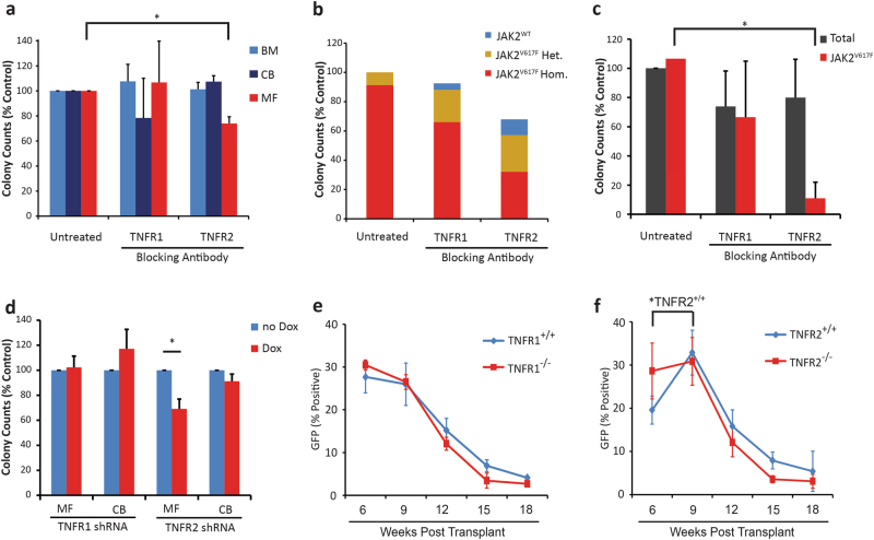
Fig. 2.
Blocking TNFR2 selectively inhibits colony formation by MF vs. normal BM CD34+ cells and mouse JAK2V617F+ compared to JAK2V617F− progenitor cells. a Human CD34+ cells from MF blood (n = 4), normal BM (n = 3) or CB (n = 3) were treated with TNFR BAs at 10 μg/mL for 72 h in liquid culture, then plated in clonogenic assays with continued treatment. TNFR2 inhibition reduced colony numbers in MF samples without affecting normal BM or CB control samples. b In MF samples, the JAK2 genotype was analyzed for each colony. Of the four samples tested, three were found to have 100% JAK2V617F allele burden in all conditions tested, while one sample with a mixed JAK2 genotype had a reduction in JAK2V617F+ colonies and increase in JAK2WT colonies with TNFR2 inhibition and to a lesser degree with TNFR1 inhibition. c Lin−Kit+ BM cells from MPN mice (n = 3) were treated in liquid culture with TNFR BAs at 10 μg/mL for 72 h, then plated in clonogenic assays with continued treatment. Colonies were enumerated after 10 days and isolated colonies genotyped for JAK2 status by analyzing GFP expression. TNFR2 inhibition selectively reduced colony formation of JAK2V617F+ cells without affecting total colony numbers, while TNFR1 inhibition had no effect. d MF (n = 4) and CB (n = 4) CD34+ cells were infected with doxycycline-inducible TNFR shRNAs, in liquid culture ±200 ng/mL doxycycline for 96 h and then plated in clonogenic assays with continued treatment. Induction of the TNFR2 shRNAs reduced colony formation in MF samples without affecting normal controls, while induction with a TNFR1 shRNA had no effect. e, f BM cells from 5-FU-treated CD45.1 and e TNFR1−/− or f TNFR2−/− CD45.2 mice were infected with MSCV-IRES-JAK2V617F-GFP retrovirus. Equal numbers of wild type and null GFP+ cells were injected into lethally irradiated TNFR+/+ recipients (n = 8 per group). TNFR2+/+ GFP+ cells increased over TNFR2−/− GFP+ cells, while TNFR1+/+ GFP+ cells remained at the same level as TNFR1−/− cells. However, GFP+ engraftment was not maintained. *P < 0.05

