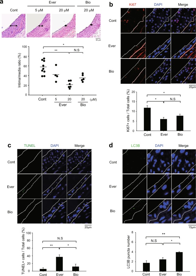
Figure 4.
Different effects of rapamycin analogues on neointimal hyperplasia in injured carotid arteries. (a) Representative histological images of balloon-injured rat carotid arteries. The injured arteries were treated with control vehicle (0.1% dimethyl sulfoxide in PBS, Cont) or everolimus (5 and 20 μM in PBS, Ever) or biolimus (20 μM in PBS, Bio) for 15 min. Data in the graph are means ± SEM of intima-to-media ratio (n = 5–6 rats per group, *P < 0.05, **P < 0.001 with one-way ANOVA). N.S., not significant (b,c) Immunofluorecence staining for Ki67 (b) or TUNEL assay (c) in the tissue sections of injured carotid arteries. Data in the graph are means ± SEM of the percentages of Ki67+ cells (b) or TUNEL+ apoptotic cells (c) versus DAPI-labelled total cells in the lesion (dashed lines). DAPI indicates nuclei. (n = 3, *P < 0.05, **P < 0.01 with Student’s t-test). N.S., not significant (d) Immunofluorescence staining of LC3B in the tissue sections of injured carotid arteries. The number of LC3B punta was counted in the marked lesions (dashed line). Data in the graph show means ± SD of total number of LC3B puncta (n = 3, *P < 0.05, **P < 0.01 with Student’s t-test). N.S., not significant. Representative images are shown.

