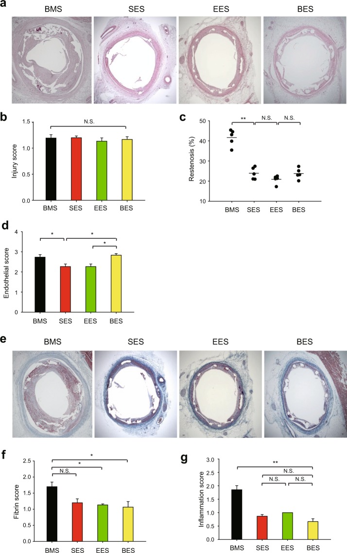
Figure 5.
Histopathological analysis in the porcine coronary arteries with stent implantation. (a) Restenosis level in the cross-section of porcine coronary arteries implanted with rapalogue-coated metal stent. Representative HE images are shown. (b) Injury score representing similar level of vascular injury by stent implantation. (c) The ratio of neointimal area versus internal elastic luminal area was calculated as described in the Materials and Methods. Data in the graph show means ± SEM of the percent of restenosis area. (d) Endothelial score was measured in the tissue sections of porcine coronary arteries implanted with rapalogue-coated metal stent. (e,f) Fibrin score was measured by Carstairs’s fibrin staining in the tissue sections of porcine coronary arteries implanted with rapalogue-coated metal stent. Representative fibrin staining images are shown (e). (g) Inflammation score was measured in the tissue sections of porcine coronary arteries implanted with rapalogue-coated metal stent. Data in graphs b,c,d,f, and g show means ± SEM of the measured scores (n = 5, *P < 0.05, **P < 0.001 with one-way ANOVA). N.S., not significant. Bare metal stent (BMS) is used as the uncoated stent control.

