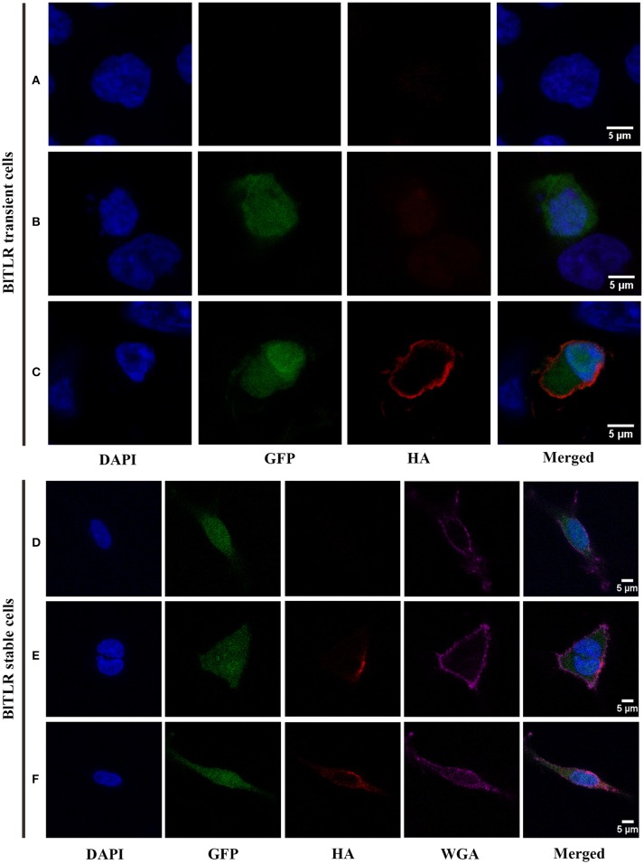
Figure 5.
Subcellular localization of BlTLR in HEK293 cells. Confocal images showing HEK293 cells transiently transfected (A–C) or stably transfected (D–F) with BlTLR. (A) Not transfected cells; (B) Cells transfected with BlTLR and non-permeabilized; (C) Cells transfected with BlTLR and permeabilized with 0.2% Triton X-100. (D) BlTLR stable cells not permeabilized; (E) BlTLR stable cells permeabilized using freeze and thaw protocol; (F) BlTLR stable cells permeabilized with 0.1% Tween-20. Nuclei are stained with DAPI (in blue). Transfected cells were GFP labeled (in green). BlTLR was detected with anti-HA antibody and AF555-conjugated anti-mouse IgG (in red). Plasma membrane was stained with WGA AF647 conjugated (in purple). Figures were analyzed with Fiji software.

