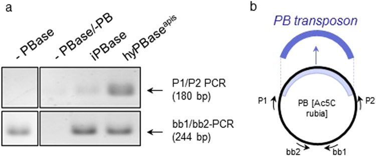
Figure 2.
The excision of PB-transposon in response to the transposases iPBase and hyPBaseapis in Sf21 cells. (a) Semi-quantitative analysis of the amount of excised transposon using PCR. A total of 106 cells were transfected with 1 µg of the PB[Ac5C rubia] plasmid23 and 1 µg of pIZ/V5-His PBase plasmid expressing either iPBase or hyPBaseapis. We isolated plasmid DNA and detected PCR fragments only if the transposon was excised (P1/P2 PCR). The PCR reactions of the different treatments were semi-quantitatively standardized for the amount of transfected plasmid. To do so, we adjusted the template’s volumes in the PCR reactions so that they produced similar strong PCR products using the bb1 and bb2 primers. Fragments were resolved via gel electrophoresis and were visualized using ethidium bromide (figure was assembled from the same gel). (b) Schematic presentation of the targets of the two PCR reactions in the PB[Ac5C rubia] plasmid.

