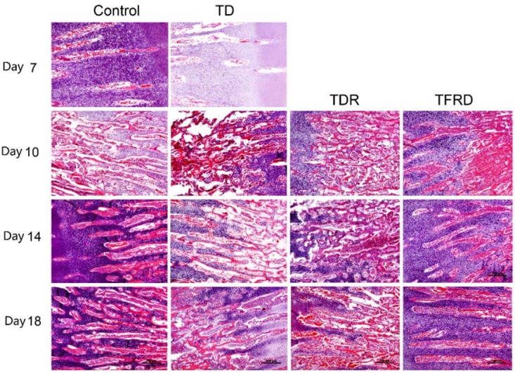
FIGURE 6.

The histopathological micrograph of H&E staining was used to describe the growth and morphological changes of blood vessels in the tibial growth plate on various days 7, 10, 14, and 18. Compared to TD and TDR group, TFRD group showed better results. The difference was not significant between control and TFRD groups.
