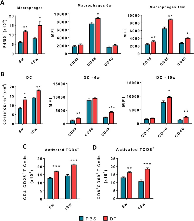
Figure 4.
DT treatment of DEREG mice in ongoing PCM increases the number and activation of macrophages, DCs and T cells that migrate to the site of lesion. The phenotypic analysis of macrophages (CD11c+F4/80+) and dendritic cells (CD11b+CD11c+) cells and some activation markers (CD80, CD86, CD40) of lung infiltrating leukocytes from DT-treated and control (PBS treated) DEREG mice was performed at weeks 6 and 10 week after P.brasiliensis infection (A,B). The lung cells were obtained as described in Material and Methods and labeled with antibodies conjugated to different fluorochromes. The lung infiltrating leukocytes were gated by FSC/SSC analysis. The cells were gated for CD11c/CD11b expression and then the CD11b cells were characterized for F4/80 expression. Macrophages and DC markers (CD80, CD86 and CD40) were then assessed in both cell subpopulations (A,B). For T cells phenotyping the lung infiltrating leukocytes were gated by FSC/SSC analysis and then gated for CD4+ or CD8+ expression. The expression of CD25 and CD69 activation markers were then evaluated in CD4+ and CD8+ T cells, respectively. One hundred thousand cells were acquired on FACS CANTO II and subsequently analyzed by FlowJo software. Data are expressed as means ± SEM of three independent experiments using 5 mice per group (*p < 0.05, **p < 0.005 and ***p < 0.001).

