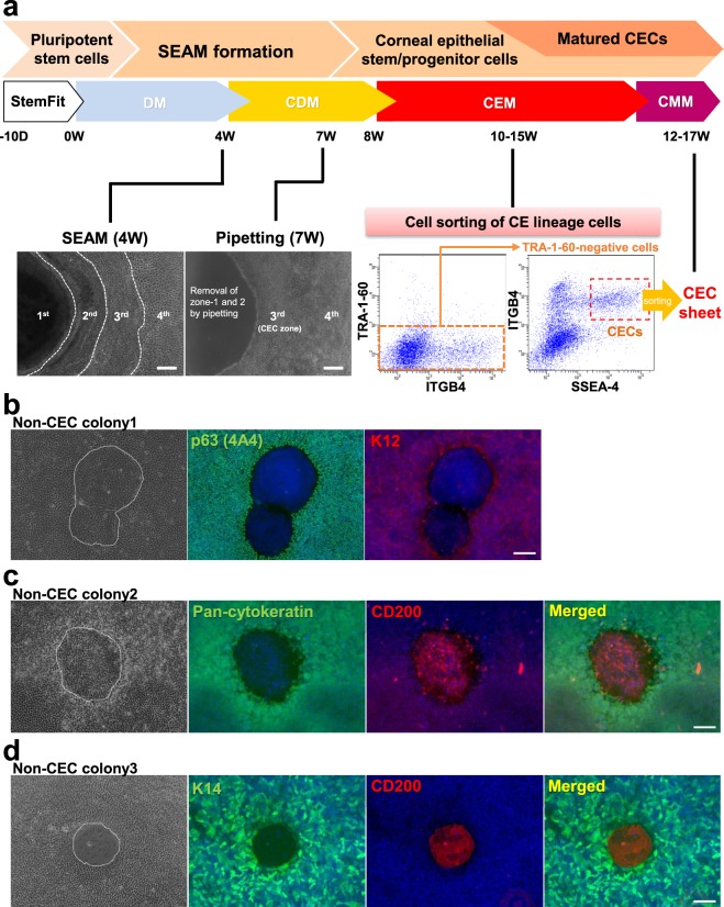
Figure 1.
SEAM formation and the insolation of human iPSC-derived CE lineage cells. (a) Schematic representation of the procedure used for the generation of a SEAM from human iPSCs and the subsequent isolation of CE lineage cells. CECs appeared in zone 3 of the SEAM at 3 to 4 weeks after the initiation of differentiation. Non-epithelial cellular zones 1 and 2 were removed by pipetting at week 7. At weeks 10 to 15, iPSC-derived CE lineage cells were isolated by cell sorting using anti-SSEA-4, anti-ITGB4, and anti-TRA-1-60 antibodies. Bars, 200 μm. (b) Representative immunostaining images showing how the expression of corneal epithelial markers p63 (green) and K12 (red) in non-CEC colony (no. 1) emerged in the iPSC-derived CEC sheets. (c,d) Representative immunostaining images of epithelial cell markers, pan-cytokeratin or K14 (green) and CD200 (red), in non-CEC colonies (nos 2, 3) in the iPSC-derived CEC sheets. Nuclei, blue. Scale bars, 50 μm.

