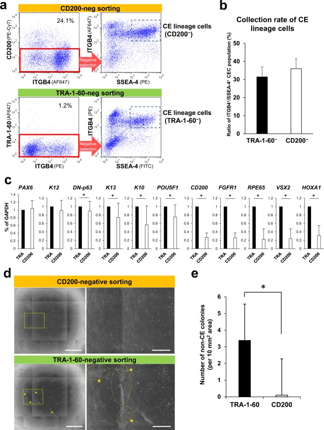
Figure 3.
Isolation of human iPSC-derived CE lineage cells by CD200-negative sorting. (a) Flow cytometric analyses of SSEA-4, ITGB4, and CD200 (upper panels) or TRA-1-60 (lower panels) expression by differentiated iPSCs at weeks 10–15 of differentiation. ITGB4+/SSEA-4+ cells were considered to be CE lineage cells obtained from CD200- or TRA-1-60-negative populations, respectively. (b) Collection rate of iPSC-derived CE lineage cell population (ITGB4+/SSEA-4+) obtained by negative sorting procedures. Error bars, SD (n = 6). (c) Expression of corneal and non-target cell-related genes by the isolated iPSC-derived CE-like cells. Relative expression levels were set as 1 in TRA-1-60-negative samples (TRA). Error bars, SD (n = 9); *p < 0.05. (d) Representative phase-contrast images showing the cultivated iPSC-derived CE-like cells isolated by CD200- (upper panels) or TRA-1-60-negative sorting (lower panels). Right: magnified areas shown in the left panels. Asterisks: non-target colonies. Scale bars: 2 mm (left), 500 μm (right). (e) Numbers of non-target colonies in the cultivated iPSC-derived CE-like cells. Error bars, SD (n = 4); *p < 0.05.

