Figure 4.
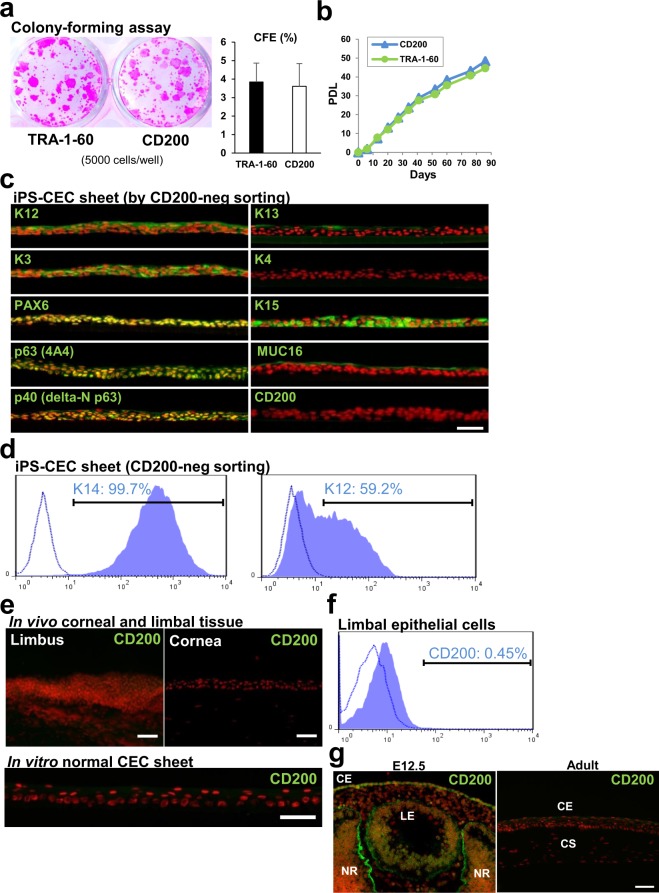
CFE of human iPSC-derived CE lineage cells, and CD200 expression in corneal tissue. (a) CFA analysis performed using iPSC-derived CE lineage cells isolated by CD200- or TRA-1-60-negative sorting (5000 cells/well). Right, CFEs of both samples. Error bars, SD (n = 4). (b) Serial cell passaging assay for iPSC-derived CE lineage cells isolated by CD200- or TRA-1-60-negative sorting. PDL; Population doubling level. (c) Representative immunostaining images showing corneal-related maker and CD200 expression (green) by iPSC-derived CEC sheets obtained by CD200-negative sorting. Nuclei, red. Scale bar, 50 μm. (d) Flow cytometric analysis of K14 and K12 expression by iPSC-derived CE lineage cells obtained by CD200-negative sorting. (e) Representative CD200 immunostaining images showing its non-expression by the corneal and limbal tissues as well as cultivated limbal epithelial cell sheet derived from human limbal tissue. Nuclei, red. Scale bars, 50 μm. (f) Flow cytometric analysis of CD200 expression by limbal epithelial cells. (g) Immunostaining of CD200 (green) in murine embryonic (E12.5) and adult eyes. Nuclei, red. CE; Corneal epithelium, CS; Corneal stroma, LE; Lens, NR; Neuro-retina. Scale bar, 50 μm.

