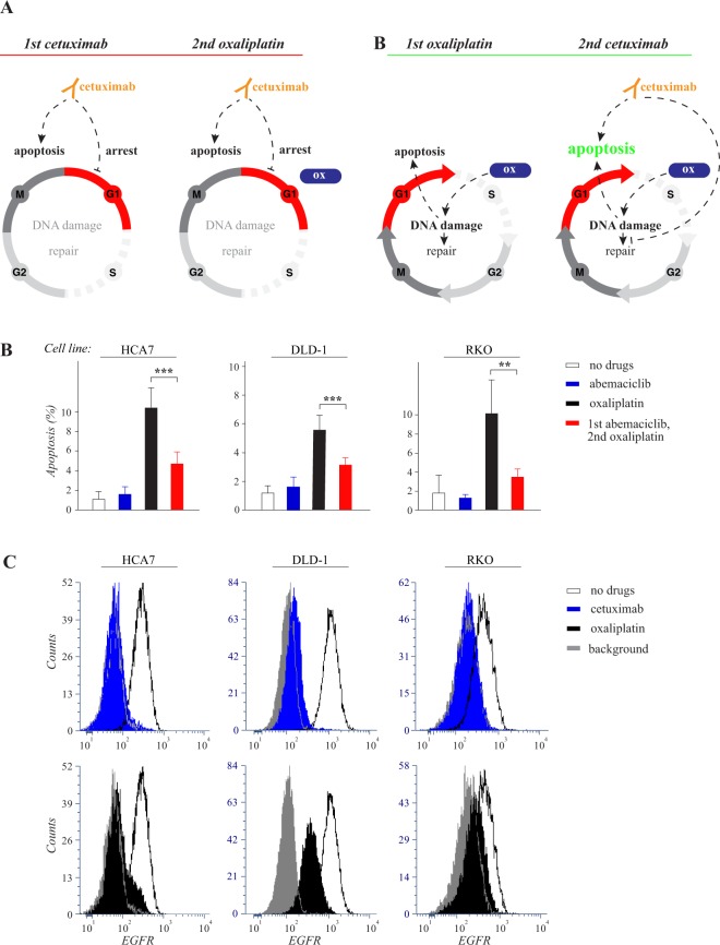
Figure 5.
Oxaliplatin is less efficient in G1-arrested cells but induces EGFR downregulation. (A) Schematic representation of hypothetical cellular responses to the opposite sequences of cetuximab and oxaliplatin. Left side: When cetuximab is administered first, it promotes arrest in the G1 phase of the cell cycle and stimulates apoptosis by blocking EGFR-activated survival pathways. The subsequent administration of oxaliplatin (ox) is largely ineffective due to the G1-arrest. Right side: When oxaliplatin is administered first, it actively induces DNA damage provoking apoptosis, as well as DNA repair that partially compensates for the damage. In the absence of cetuximab, the cell cycle is not arrested at G1 allowing for full activity of oxaliplatin. However, oxaliplatin-induced DNA damage itself does eventually cause cell cycle arrest by activating checkpoints in the S and G2 phases (not indicated in the figure). When cetuximab is then added after oxaliplatin, apoptosis is enhanced compared to all other regimens, as the apoptotic effect of both drugs are active, and also, as cetuximab may be suppressing the DNA repair mechanisms caused by oxaliplatin-induced DNA damage. (B) HCA7, DLD-1 and RKO cells were arrested in G1 by 8 hour treatment with or without 3 µM of the CDK4/6 inhibitor abemaciclib followed by 18 hour treatment with or without 50 μM oxaliplatin. Apoptosis was measured by annexin V analysis. Mean +/− SD is shown for three independent experiments. **P < 0.01; ***P < 0.001. (C) Flow cytometry analysis of cell surface EGFR after 24 hour treatment with or without 10 μg/ml cetuximab or 50 μM oxaliplatin.

