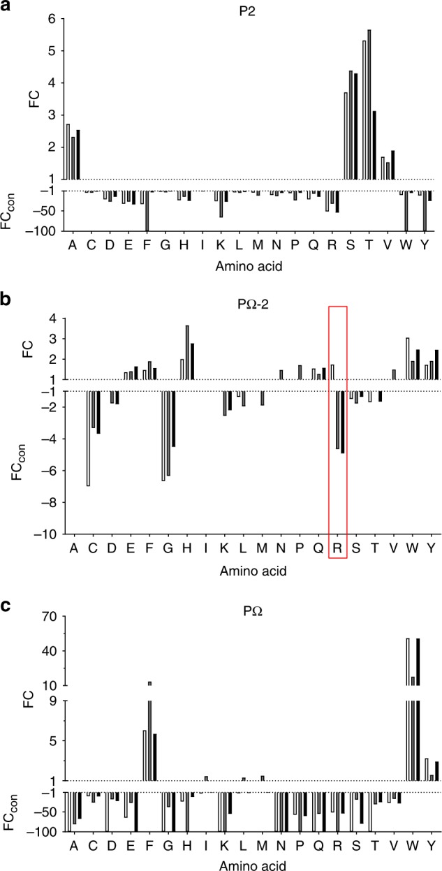
Fig. 2.

Enrichment/diminution of residues at P2, PΩ-2 and PΩ of HLA-B57 family ligands distinguish primary and secondary anchor sites. The enrichment of specific residues at P2 (a), PΩ-2 (b) and PΩ (c) of all 9 residue peptide binders of HLA-B*57:01 (clear bars, 912 peptides), HLA-B*57:03 (grey bars, 1155 peptides) and HLA-B*58:01 (black bars, 960 peptides) identified by LC-MS/MS relative to amino acid frequencies in the human proteome. Deviations in prevalence from the human proteome are depicted as either a fold change (FC, for amino acids at higher prevalence than in the human proteome) or a converted FC (FCcon = −1/FC, for amino acids present at lower prevalence than in the human proteome), as determined using iceLogo v1.2 stand-alone software31 using the static reference method (reference set Homo sapiens Swiss-Prot means, p < 0.05). FC > 1 indicates enrichment, FCcon < −1 indicates disfavoured residues, −100 indicates absence. In b the unique enrichment of Arg(R) at PΩ-2 by HLA-B*57:01 is highlighted by the red box
