

Abstract
The anatomy of the lower lateral cartilage varies according to the ethnicity of the patient. Considering that the manipulation of the lower lateral cartilage has become more prevalent in Indian rhinoplasties, understanding the comprehensive anatomy is of utmost importance. The aim of this descriptive study was to evaluate the anthropometric and morphological variations of the lower lateral cartilage in Indian noses and to compare this data from studies of various ethnic groups. Seventy lower lateral cartilages of thirty-five patients of Indian origin who underwent primary open rhinoplasty were dissected and assessed intraoperatively. There was no previous history of nasal trauma. The medial, middle and lateral distances from the caudal border of the alar cartilage to the alar rim were measured. Morphology of the cartilage was assessed. The results were analysed and comparison were made between the genders and various ethnic groups. A statistically significant difference (p < 0.05) was observed in terms of length, width, distance from the alar rim, compared to the other study. Convex type (56%) of lateral crura was most commonly seen, with no significant gender difference. This study highlights the anatomical differences among various ethnic groups and stresses the need to be aware of the complexities of the anatomical aspect of the cartilage, to avoid complications and provide acceptable aesthetic result to the patient.
Keywords: Lower lateral cartilage, Lateral crus, Indian rhinoplasty, Primary open rhinoplasty
Introduction
Ancient Indian literature is rife with rich information and valuable knowledge about the country’s culture, religion and medical science. One of the very important medical contribution for which Indians are credited is rhinoplasty.
The origin of reconstruction of nose can be traced to the early days of recorded history and is unique culture heritage of India. The nose is the important and prominent organ of the body which carries great emotional and social significance. Any deformity of the nose affects the person physically, socially and psychologically. The first description of nasal reconstruction is mentioned in the extensive work of Susruta in Susruta Samhita in as early as sixth century B.C. [1].
The external nose consists of four major areas—Bony pyramid, Cartilaginous pyramid, Lobule and soft tissue areas. The bony pyramid, Cartilaginous pyramid, and lobule forms one-third of the external nose and form an integrated anatomical—physiological entity. The bony pyramid consists of the nasal bones, nasal part of frontal bone and two frontal process of the maxilla. The nasal bones are thicker and narrower cranially and thinner and wider caudally and attached in midline by internasal suture and unite with nasal part of frontal bone by frontonasal suture [2].
The septolateral cartilage and two lateral membranous areas with accessory cartilages comprises the cartilaginous vault. The septolateral cartilage consists of the cartilaginous septum and upper lateral cartilages and the upper margin of the cartilages underlaps the nasal bones by 1 or 2 mm. The area where the nasal bones, the septal cartilage and the two triangular cartilages unite is called Keystone area.
The lobule is the lower third of the external nasal pyramid. It is made of two lower alar cartilage, muscle fibres, subcutaneous connective and fatty tissue and thick skin. The lower alar cartilages are horse shoe shaped cartilages that support the lobule structure. They determine the position and form of the tip, alae and columella and configuration of nares and vestibules. They can be divided into three parts—medial, intermediate and lateral crus.
The medial crura forms the columella along with the lower portion of the septal cartilage and consists of two components—the footplate segment and columellar segment. The intermediate crus consist of lobular and domal segment. The domal segment is strongly bent part between the medial and lateral crura and the two domes together make the nasal tip and are connected by interdomal ligament or Pitanguy ligament. The lateral crus are the largest component of the nasal lobule and plays a major role in defining the shape of anterior superior portion of alar sidewalls. The caudal edge parallels and provide support for only anterior half of alar rim, so, any excessive excision of the medial half of the crura can cause weakening of support of anterior alar rim [3].
The nasal tip has been the major beneficiary of the open approach since it imposes the tip structures in their natural undisturbed position. It also allows unparalleled diagnosis of the deformities and asymmetries as well as facilitate precise surgical manipulation of the lobule. It allows the correction of minor irregularities, thus adding extra surgical finesse.
The concept of ethnic nose has evolved over time. Asia is a diverse continent including people from different ethnicities including Eastern Asia, Southeast Asia, India and Middle east [4]. Indians among them have more variability owing to the substantial climatic changes in the country. The people in North India have more Caucasian feature whereas South Indians have Australoid features and Eastern Indians have mongoloid features. This could also be explained due to the great shift that occurred thousands of years ago. The typical Indian nose lacks projection, the osteocartilagenous framework is broad, the tip is ill—defined and lobule is broad [5]. Size, shape and orientation of the lateral crura of the lower lateral cartilage (LCC) greatly influence the aesthetics and function of the lower one-third of the nose. As the lateral crura extend laterally from the intermediate crura, a mild concavity typically exists. Excess convexity of the lateral crura is often seen in the bulbous, boxy and “parentheses” tip deformities [6]. The common indications for rhinoplasty in Indians are ill defined nose, lack of projection, contour deformities and deviated nose. The typical Indian nose lacks projection and hence require augmentation more often than reduction [5]. The classical rhinoplasty technique described in western literature may not be fully applied to the Indian context. Also, the country boasts of largest diaspora in the world with more than 15.6 million people of Indian origin living abroad. Many of the said population, owing to the current trend of medical tourism and consciousness of the cosmesis of nose because of awareness and media, are seeking aesthetic surgery in different parts of the world.
Considering that the manipulation of the alar cartilage has become more prevalent in Indian rhinoplasties, understanding the detailed anatomy is of utmost importance for the surgeons in the country as well as worldwide. There are six major ethnic groups that have been anatomically described in the rhinoplasty literature: Caucasians, Asians, African, Mediterranean, Middle eastern and Latin American. To date as per the available literature and references, no anatomic or anthropometric study of the lower lateral cartilages has been performed in India, even though rhinoplasty is one of the most common and evolving field of surgery in the country.
Aim
The aim of this descriptive study was to evaluate the anthropometric and morphological variations of the lower lateral cartilage in Indian noses and to compare this data from studies of various ethnic groups.
Materials and Methods
Study Design
Descriptive study.
Sample Size
A total of 35 patients underwent open rhinoplasty in which 70 alar cartilages were measured.
Inclusion Criteria
Patients above the age of 18 years.
Exclusion Criteria
Patient who have undergone previous nasal surgeries.
Patients having any diseases of the nose which are causing destruction or deformities of the nose.
Patients having obvious history of trauma or injury to nose.
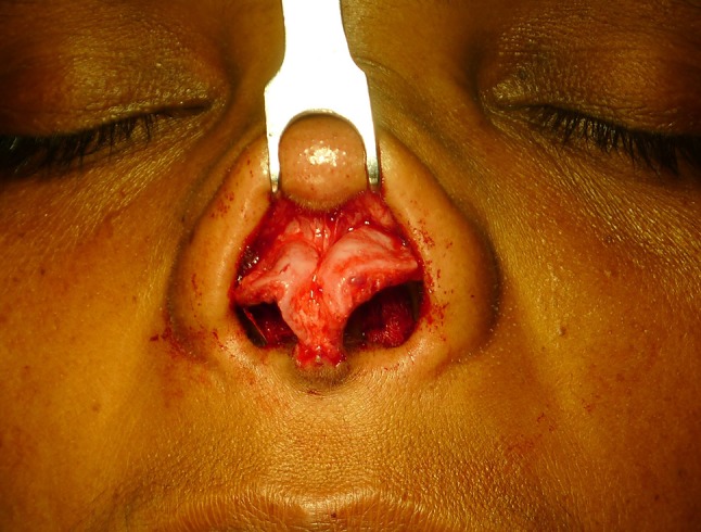
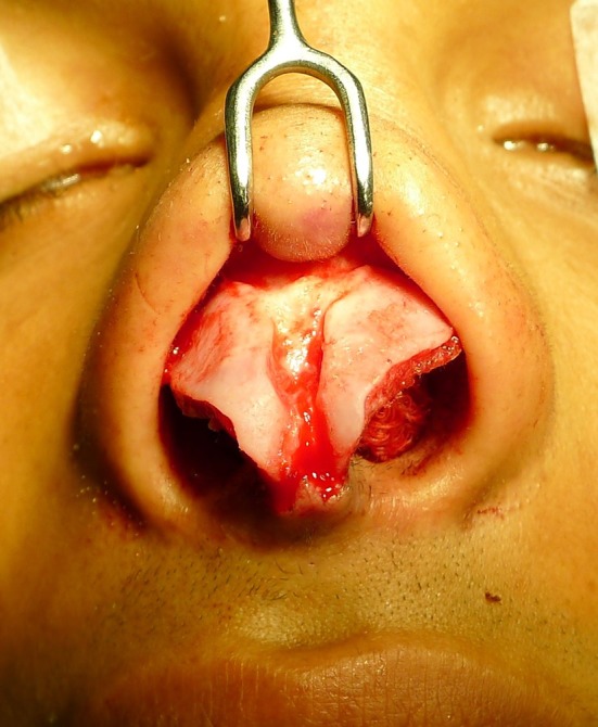
Patients details and history were obtained. Detailed clinical examination was done. Informed consent was obtained. Preoperative pictures of the nose of the patient was taken methodically. A standard open rhinoplasty approach was used. A mid columellar incision was given followed by marginal incision. Lower lateral cartilages were completely dissected and accurate measurements were taken. The parameters included were the greatest length, width, thickness of each lateral crus and the distances of the caudal border of the lateral crus from the alar rim points at different levels representing the medial, middle, and lateral portions. Morphology of the lateral crus was observed based on which they were divided into types such as concave, convex, concave–convex, convex–concave, and flat. Measurements were taken with electronic digital Vernier caliper indicating millimetres up to a decimal of 0.00 mm. A single surgeon performed all the surgeries included in the study (Fig. 1).
Fig. 1.

Illustration of the areas measured. Length and width of the lateral crus and distance of the lower border of lateral crus to the alar rim
Results were evaluated by means and standard deviations. Male and female scores were compared by t test.
Results
Twenty-one patients (60%) were male and 14 patients (40%) were females.
Table 1 shows the results of the length, width and thickness of the lateral crura. The most common contour of the lateral crus was convex (56%) of LLCs followed by Flat (16%), Convex–Concave (14%) and Concave (14%). Right and left lateral crura mean length was 15.32 ± 2.20 and 15.28 ± 2.28 mm respectively, width was 9.97 ± 1.47 and 9.53 ± 1.61 mm respectively and thickness was 0.90 ± 0.25 and 0.95 ± 0.25 mm respectively showed no significant difference between genders.
Table 1.
Comparison of size of the lateral crus in different races
| Zelnik and Gingrass (Caucasians) [7] | Dhong et al. (Koreans) [8] | Farhvash et al. (Persian) [9] | Ofodile et al. (African-American) [10] | Present study (Indians) | |
|---|---|---|---|---|---|
| Length | 18–27 (23.1) | 17.2 ± 2.2 Male: 19.7 ± 3.2 Female: 16.8 ± 1.1 |
Right—23.4 ± 2.7 Left 23.1 ± 2.5 |
9–25 (18) | Males: Right 15.86 ± 2.43 Left 15.46 ± 2.42 Female: Right 14.51 ± 1.56 Left 15.01 ± 2.11 |
| Width | 10–15 (12.6) | 10.0 ± 1.3 Male: 10.5 ± 1.5 Female: 9.8 ± 1.1 |
Males: Right 11.2 ± 1.4 Left 10.2 ± 0.7 Females: Right 11.2 ± 1.4 Left 10.3 ± 0.8 |
8—14 (12) | Males: Right 10.03 ± 1.22 Left 9.44 ± 1.63 Female: Right 9.87 ± 1.82 Left 9.67 ± 1.64 |
| Thickness | Males: 0.55 ± 0.05 Females: 0.54 ± 0.15 |
Males: Right 0.9 ± 0.16 Left 0.85 ± 0.16 Female: Right 0.8 ± 0.14 Left 0.8 ± 0.15 |
Males: Right 0.93 ± 0.19 Left 0.98 ± 0.25 Female: Right 0.86 ± 0.31 Left 0.90 ± 0.24 |
The distance of the lateral crura from the alar rim medially was 2.77 ± 0.77 mm, at the middle was 4.62 ± 1.04 mm and laterally was 7.00 ± 1.35 mm (Tables 2 and 3).
Table 2.
Comparison of distance of the lateral crus in different races
| Zelnik and Gingrass (Caucasians) [7] | Dhong et al. (Koreans) [8] | Farhvash et al. (Persian) [9] | Ofodile et al. (African-American) [10] | Present study (Indians) | |
|---|---|---|---|---|---|
| Medial | 4–8 (6) | Male: 5.7 ± 1.1 Female: 5.8 ± 1.1 |
Male: 5–8 (6) Female: 4–7 (5) |
5—9 (6) | Males: Right 2.89 ± 0.68 Left 2.86 ± 0.80 Female: Right 2.57 ± 1.06 Left 2.65 ± 0.50 |
| Middle | 3–7 (5) | Male: 7.4 ± 1.4 Female: 6.5 ± 1.1 |
Male: 6–8 (7) Female: 5–7 (6) |
4–8 (6) | Males: Right 4.70 ± 0.82 Left 4.64 ± 1.11 Female: Right 4.70 ± 1.16 Left 4.40 ± 1.15 |
| Lateral | 9–19 (13) | Male: 12.3 ± 2.1 Female: 11.5 ± 1.5 |
Male: 10–22 (14) Female: 8–19 (12) |
10–15 (11) | Males: Right 7.14 ± 1.29 Left 6.97 ± 1.28 Female: Right 6.97 ± 1.72 Left 6.83 ± 1.27 |
Table 3.
Comparison of the morphological types of the Alar Cartilage
Discussion
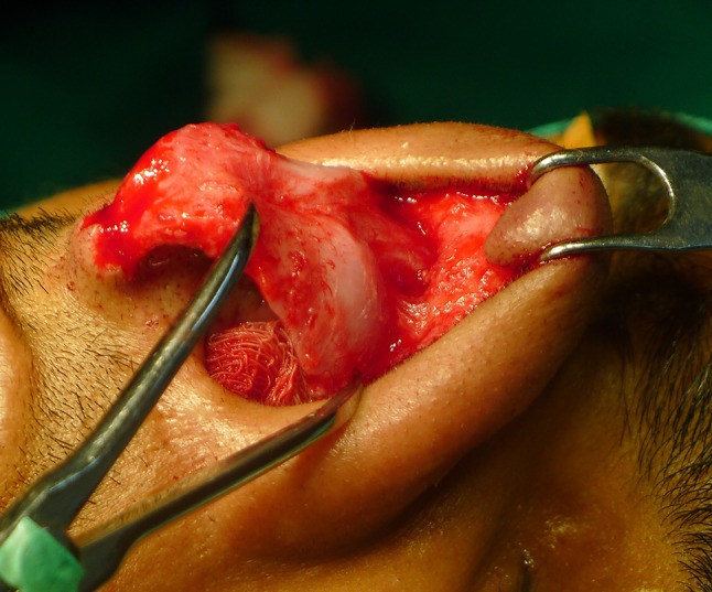
Our study was first of its kind performed in India and had a relatively large number. Seventy alar cartilages were measured and analysed. The dimensions of the cartilage were obtained by meticulous dissection of the soft tissues over the cartilage intraoperatively. This excludes the drawbacks of the measurement taken on cadavers as there will be some amount of tissue loss and deformity of the cartilage due to preservation techniques of the cadaver (Figs. 2, 3, 4, 5, 6 and 7).
Fig. 2.

Concave type
Fig. 3.
Convex concave type
Fig. 4.

Convex type (frontal)
Fig. 5.

Convex type (lateral)
Fig. 6.
Flat type
Fig. 7.
Intraoperative measurement with castroveijo caliper
Though, we were not able to find a significant correlation (p < 0.05) between males and females within our population in terms of length, width and thickness of the cartilage, in general the female LLCs was smaller, narrower and thinner. In a study on Persian lower lateral cartilages, there was a significant difference in terms of width between the two [9]. When comparison was made between the length and width of the cartilages with that of the Persians, the cartilage in Indians was smaller and leaner. This was significant in both the genders (p < 0.05). When comparison was made with Asians, the cartilage in Indians was smaller and slender. This, though not expected, could be explained by the presence of increased skin thickness in Indians. (p < 0.001). The thickness of the lateral crus was more in males than females, which was same as Korean study [8].
The mean distance from the lower border of the lateral crus to alar rim point in Koreans was 5.7, 7.4 and 12.3 mm whereas in our study it was 2.77, 4.62 and 7.00 mm. This proves that the LLC in our population are placed more caudally and hence knowing the exact dimensions is important while taking marginal incision for open rhinoplasty approach [8].
The Lower lateral cartilages in our study appeared to be smaller and narrower than those of other ethnic groups.
The morphology of the lateral crus was based on the morphological types based on Zelnik et al. [7] and Ofodile and James [10] into convex, concave, convex–concave, concave–convex and flat. The most common morphology was convex (56%), Concave (14%), Flat (16%) and convex concave (14%). Convex–Concave type of cartilage was commonest in Koreans (30%) and African Americans (50%) whereas it was Convex type in Persians (89.9%). Concave type of cartilage was not seen in females in the study which are usually expressed clinically as alar depression. It was also found that concave lateral crura provides a more convex domal area, requiring limited manipulation at the tip. Excessive convexity of the lateral crura is often seen in bulbous and boxy tip deformities.
Bulbous tip correction requires excision of the cephalic portion of the large alar cartilage. As the cartilage is narrow in Indians, excision of the cephalic portion should be limited to avoid alar collapse. The measurements showed the lateral aspect of the alar cartilage to be closer to the nostril than other ethnic groups and care should be taken to avoid visible scarring or transecting the lower lateral cartilages.
As we do not operate on ethnic groups but operate on individuals, an illustrative understanding of the anatomy of the lower lateral cartilages in different ethnic groups, can prevent secondary rhinoplasties. Pertaining to the nasal architecture, there are subtle characteristics inherent to each race. As we know, India has a sizeable diaspora, thus this study could act as a reference point for non-resident Indians undergoing rhinoplasty in a foreign land.
Conclusion
We evaluated the anthropometry and morphology of the alar cartilage in Indian patients and compared it with those reported in studies of other ethnic groups. Our findings show that Indian anthropometric measurements differ from those in Asians, Caucasians, and African Americans. This stresses the need of the aesthetic surgeon of the nose, all over the globe, to be aware and understand the intricacies of applied anatomical aspect of the Indian alar cartilage architecture to avoid complications and to provide acceptable aesthetic and functional result to the patients. Thus, our study analyses these variations and gives accurate measurement of the alar cartilage, to perform acceptable rhinoplasty in Indian population worldwide.
Conflict of interest
No Conflict of Interest.
Ethical Clearance
Obtained from the Institution's Ethical Clearance Comittee.
References
- 1.Sood V (ed) (2002) History of rhinoplasty. In: Corrective rhinoplasty, 2nd edn. CBS Publisher and distributors, pp 1–8
- 2.Huizing EH, Chairman F, De Groot JAM, Assistant F (eds) (2015) Functional reconstructive nasal surgery. In: Surgical anatomy, 2nd edn. Thieme, pp 2–28
- 3.Oneal RM, Beil RJ (2013) Surgical anatomy of the nose. In: M Shiffman, A Giuseppe (eds) Advanced aesthetic rhinoplasty: art, science, and new clinical techniques, Springer, pp 33–60
- 4.Jang YJ (2013) Asian rhinoplasty. In: M Shiffman, A Giuseppe (eds) Advanced aesthetic rhinoplasty: art, science, and new clinical techniques, Springer, pp 163–173
- 5.Bhat U, Patel B. Primary rhinoplasty: an Indian perspective. Indian J Plast Surg. 2008;41:S9–S19. [PMC free article] [PubMed] [Google Scholar]
- 6.Rohrich RJ, Adams WP. The boxy nasal tip: classification and management based on alar cartilage suturing techniques. Plast Reconstr Surg. 2001;107(7):1849–1863–1868. doi: 10.1097/00006534-200106000-00034. [DOI] [PubMed] [Google Scholar]
- 7.Zelnik J, Gingrass RP. Anatomy of the alar cartilage. Plast Reconstr Surg. 1979;64:650–653. doi: 10.1097/00006534-197964050-00008. [DOI] [PubMed] [Google Scholar]
- 8.Dhong E-S, Han S-K, Lee C-H, Yoon E-S, Kim W-K. Anthropometric study of alar cartilage in Asians. Ann Plast Surg. 2002;48(4):386–391. doi: 10.1097/00000637-200204000-00009. [DOI] [PubMed] [Google Scholar]
- 9.Farahvash MR, Ebrahimi A, Farahvash B, Farahvash Y. Anatomic and anthropometric analysis of 72 lower lateral nasal cartilages from fresh Persian (Iranian) cadavers. Aesthet Surg J. 2012;32(4):447–453. doi: 10.1177/1090820X12442084. [DOI] [PubMed] [Google Scholar]
- 10.Ofodile FA, James EA. Anatomy of alar cartilages in blacks. Plast Reconstr Surg. 1997;100(3):699–703. doi: 10.1097/00006534-199709000-00026. [DOI] [PubMed] [Google Scholar]