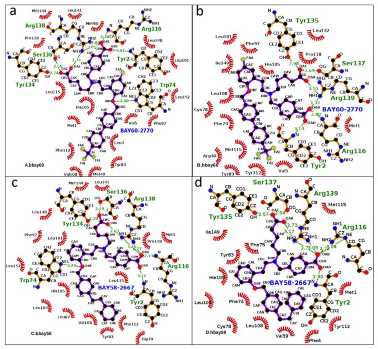
Figure 3.
From a to d, 2D graphical outlay of binding modes of sGC activators bound in the binding pockets of human and bacterial H-NOX domains (bbay60, hbay60, bbay58, hbay58). The energy minimized starting structures (hbay58 and hbay60) along with bbay58 and bbay60 (crystal structures) were subjected to MD production phase. Green dotted lines represent Hydrogen bonds whereas hydrophobic interactions are shown by red archs, the ligand atoms are shown in blue and black ball and stick model whereas protein atoms are shown in brown (stick) and black (ball). Aliphatic and aromatic carboxyl groups atoms of activators (OAA, AAB, OAC, OAD) exhibited prominent hydrogen bond interaction with functionally critical residues such as Y2, R116, Y135, S137, R139 in hH-NOX andY2, W74, R116, Y134, S136, R138 in bH-NOX.

