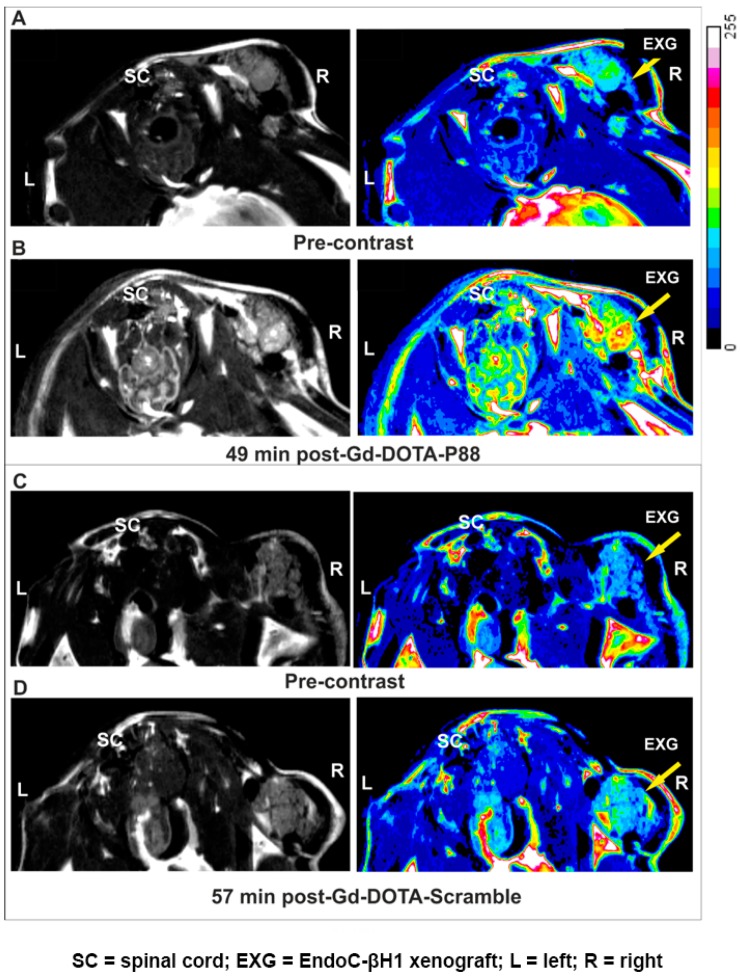
Figure 4.
Non-invasive MRI of EndoC-βH1 xenograft bearing-mice using Gd-DOTA-P88 and Gd-DOTA-Scramble. (A–D) Representative MR images of mice bearing EndoC-βH1 transplants. Pre-contrast images (A,C) were acquired before injection of CAs and the post-contrast images were obtained 50 min (B,D) after i.v. administration of 0.1 mmoL Gd/kg b.w. of Gd-DOTA-P88 (B) or Gd-DOTA-Scramble (D). Mice were implanted with EndoC-βH1 grafts in transplantation rings in the right hind leg and vehicle transplantation rings in the left hind leg. The images are displayed in grey-scale (left row) and in pseudo-coloring overlay (right row). The images are representative of 4–6 similar experiments. Legend: SC = spinal cord; EXG = EndoC-βH1 xenograft; L = left; R = right.

