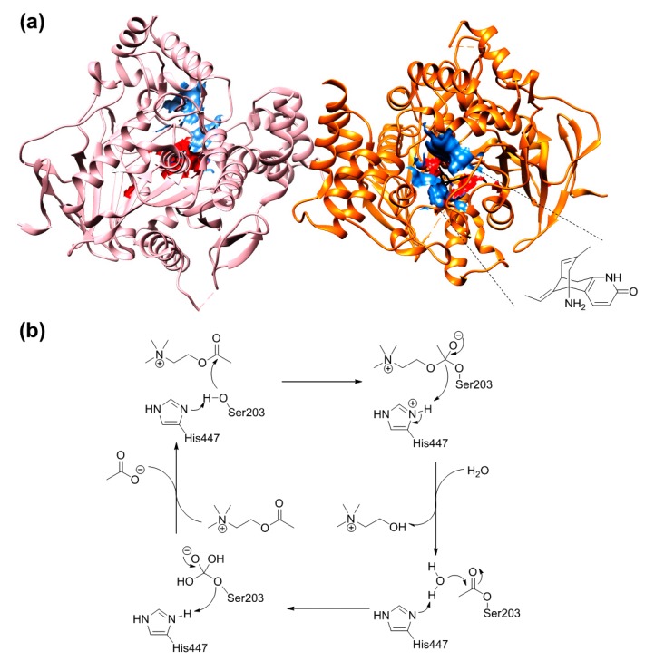
Figure 1.
The crystal structure of hAChE (PDB ID: 4EY5) in complex with huperzine A (a). Subunit A is depicted in pink ribbons whereas the B subunit is coloured in orange. Huperzine A is presented in black and its structure is highlighted with dashed lines. The anionic sub-site is described by blue spheres, while the esterase sub-area is encircled by red spheres. For the sake of clarity, hydrogen atoms were omitted. The graphic was generated using the UCSF Chimera software (Resource for Biocomputing, Visualization, and Informatics (RBVI), University of California, San Francisco, CA, USA). Biochemical mechanism of ACh hydrolysis by AChE (b).

